
FIGURE
Fig. 2
- ID
- ZDB-FIG-140304-35
- Publication
- Zhang et al., 2014 - p35 promotes the differentiation of amacrine cell subtype in the zebrafish retina under the regulation of egr1
- Other Figures
- All Figure Page
- Back to All Figure Page
Fig. 2
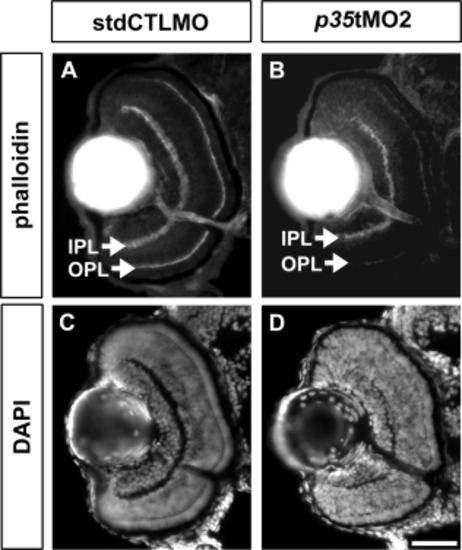
Retinal differentiation was affected by p35 knockdown at 72 hpf. The retinal histology of the p35 morphants (p35 tMO2) and controls (stdCTLMO) was analyzed at 72 hpf. At this stage, the phalloidin staining highlighted IPL and OPL with a sharp and distinct boundary in the controls (A), while these plexiform layers were thinner and irregular in the p35 morphants (B). A similar observation of the plexiform layer formation was also revealed by the DAPI nuclei staining on the same sections (C, D). IPL: inner plexiform layer; OPL: outer plexiform layer. Scale bar = 50 μm. |
Expression Data
Expression Detail
Antibody Labeling
Phenotype Data
| Fish: | |
|---|---|
| Knockdown Reagent: | |
| Observed In: | |
| Stage: | Protruding-mouth |
Phenotype Detail
Acknowledgments
This image is the copyrighted work of the attributed author or publisher, and
ZFIN has permission only to display this image to its users.
Additional permissions should be obtained from the applicable author or publisher of the image.
Full text @ Dev. Dyn.