
Fig. 2
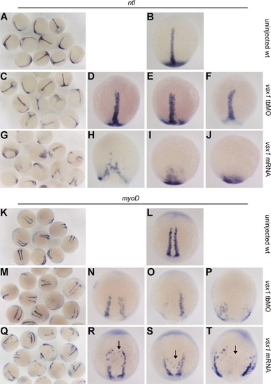
Comparison of axial and paraxial mesoderm formation among wild type, maternal Vsx1 suppressed and vsx1 overexpression embryos. (A–J) ntl marked axial mesoderm domain in uninjected wild-type control embryos (A and B), vsx1 tbMO injected embryos (C–F) and vsx1 mRNA injected embryos (G–J). (K–T) myoD marked paraxial mesoderm domain in uninjected wild-type control embryos (K and L), vsx1 tbMO injected embryos (M–P) and vsx1 mRNA injected embryos (Q–T). Note that vsx1 overexpression inhibits the convergence of paraxial mesoderm cells but has no impact on paraxial mesoderm cell specification and somite formation. Riboprobes are indicated at the top of each group of figures. All the images of single embryo are dorsal view with animal pole towards the top. |
| Genes: | |
|---|---|
| Fish: | |
| Knockdown Reagent: | |
| Anatomical Terms: | |
| Stage Range: | 5-9 somites to 10-13 somites |
| Fish: | |
|---|---|
| Knockdown Reagent: | |
| Observed In: | |
| Stage Range: | 5-9 somites to 10-13 somites |
Reprinted from Developmental Biology, 386(1), He, Y., Xu, X., Zhao, S., Ma, S., Sun, L., Liu, Z., and Luo, C., Maternal control of axial-paraxial mesoderm patterning via direct transcriptional repression in zebrafish, 96-110, Copyright (2014) with permission from Elsevier. Full text @ Dev. Biol.