
Fig. 3
- ID
- ZDB-FIG-140227-13
- Publication
- Hauptmann et al., 1996 - Complex expression of the zp-50 pou gene in the embryonic zebrafish brain is altered by overexpression of sonic hedgehog
- Other Figures
- All Figure Page
- Back to All Figure Page
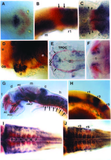
Characterization of zp-50 expression by doublelabeling. In all side and dorsal views anterior is to the left. (A-D) 2 color in situ hybridization for zp-50 (purple) and pax-2 transcripts (red). (A) Dorsal view of flat mounted bud stage embryo with zp-50 staining in the posterior diencephalon and pax-2 expression along the future midbrain/hindbrain junction. (B) Side view of the mid-/hindbrain border region of a 10 somite stage embryo. Arrows indicate a zone of overlapping zp-50 and pax-2 expression. (C) Dorsal view of 26 hpf embryo. zp-50-expressing cells in the tegmentum are separated by a gap of 2-3 cell diameters (arrowheads) from the pax-2 domain at the midbrain/hindbrain boundary. (D) Frontal view of 27 hpf embryo: pax-2 stains the optic stalks emerging next to the anteriormost position of diencephalic zp-50 expression. (E) Dorsal view of 34 hpf anterior diencephalon hybridized with zp-50 (purple). Axons are immunostained with an antibody to acetylated tubulin (red). The anteriormost zp-50 expression site is surrounded (arrows) by the axons of the postoptic commissure and its associated longitudinal tract (TPOC). (F) Dorsal view of 36 hpf telencephalon hybridized with zp-50 (purple). The zp-50-expressing cells are more medial but partially overlap with neural cells positive for the zn-12 epitope (red) projecting axons into the tract of the anterior commissure. (G) Side view of 34 hpf embryo hybridized with zp-50 (purple). Axons are stained with the acetylated tubulin antibody (red). The eyes have been removed. The small arrow in the anterior diencephalon indicates zp-50 expression located in the immediate vicinity of the junction between the telencephalic (or supraoptic) axon tract with the tract of the postoptic commissure. The white arrow shows the nucMLF which is surrounded ventrally, anteriorly and dorsally by the zp-50 expression domains. Large arrows in the area of the hindbrain indicate reticulospinal neurons in the rhombomere centers flanked by zp-50-expressing double stripes at the interrhombomeric boundaries. (H) Side view of a 5 somite embryo double labeled with zp-50 (purple) and krx-20 (red) expressed in rhombomeres r3 and r5. (I) Dorsal view of 36-40 hpf hindbrain hybridized with zp-50 (purple) and hlx-1 (red) showing similar expression domains along medial rhombomere borders. The numbers indicate the corresponding rhombomere centers. (J) Dorsal view of 36-40 hpf hindbrain doublestained for zp-50 (purple) and krx-20 (red). At this stage, krx-20 is only strongly expressed in r5 whose borders are indicated by open triangles. AC, anterior commissure; c, cerebellum; d, diencephalon; e, eye; h, hindbrain; m, mesencephalon; nMLF, nucleus of the medial longitudinal fascicle; OS, optic stalk; PC, posterior commissure; POC, postoptic commissure; r1, r3, r5, rhombomeres 1, 3 and 5; t, telencephalon; TAC, tract of the anterior commissure; TPC, tract of the posterior commissure; TPOC, tract of the postoptic commissure; TT, telencephalic (or supraoptic) tract. |