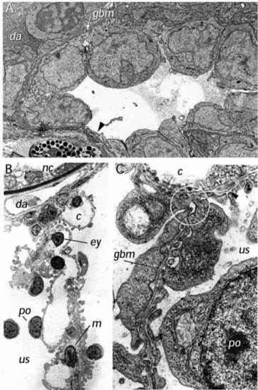
Ultrastructure of the dbb-/- glomerulus. (A) Ultrastructure of the dbb-/- glomerulus at 40 hpf. Podocytes (po) are present but appear to show less extensive podocyte foot process development at this stage compared to wild type. Areas of cellular thinning (arrow) are also evident. (B) Cross section of the glomerular septum in the mutant double bubble (3.5 dpf). The glomerular capillaries (c), mesangium (m) and podocytes (po), on either side form a medial septum between the two cysts. The urinary spaces (us) on both sides of the septum represent the lumens of the cysts. ×3,500 (C) Section of part of the dbbm468-/- glomerular septum extending between two capillaries (c), (only one capillary is shown at the top). In several places, the glomerular septum consists of two podocyte layers (po) located adjacent to each other without a capillary or a mesangial layer in between. The two epithelia are focally interconnected by cell-cell junctions and form a common glomerular basement membrane (gbm), which appears not to be continuous with the GBM of the adjacent glomerular capillaries (encircled). In some places, the GBM appears to split and rejoin (arrowhead). ×35 000. da, dorsal aorta; gbm, glomerular basement membrane; nc, notochord; e, capillary endothelial cell; ey, erythrocyte; c, capillary lumen; us, urinary space.
|
