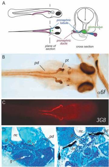
The zebrafish pronephros. (A) Diagram of the three elements of the larval pronephros at 72 hpf showing the position of glomerulus under the notochord, the pronephric tubules extending laterally to connect with the pronephric ducts which serve the function of the collecting system. (B) Horseradish peroxidase whole-mount immunostaining of a 84 h embryo with anti-Na+/K+-ATPase alpha subunit monoclonal antibody (α6F;) shows the overall anatomy of the pronephric ducts (pd) and pronephric tubules (pt), which become progressively convoluted. The glomerulus is unstained at the embryo midline. (C) The monoclonal antibody 3G8 stains the anterior half of the pronephric ducts as well as the lateral part of the pronephric tubules. (D) Cross section of a 72 h embryo showing that the glomerulus (gl), the pronephric tubule (pt) and the pronephric duct (pd) are fully formed between the gut (g) and the notochord (nc). (E) In a 6.5-day-old larva, Bowman’s space (bs) is clearly evident in the glomerulus (gl).
|
