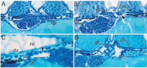
Early glomerular defects in the dbb-/- pronephros. dbbm468-/- embryos were examined at different stages of nephron development to determine the site of the primary defect. (A) Cross sections of dbbm468 pronephroi at 33 hpf reveal that the nephron primordium (np) is a double-layered sac of cells with a central lumen similar to wild-type pronephroi at this stage. (B) In 40 hpf mutant pronephroi, the distinct basement membrane separating glomerular and tubule domains is not seen. Instead, the forming glomerulus appears distended with fluid and cell-free areas (arrow) are evident at the junction of the glomerulus and tubule. (C) Longitudinal sections through the mutant glomerulus shows a distension of the glomerular lumen, flattening of glomerular cells and a thinning of cells associated with the glomerular basement membrane (arrow). (D) At 2.5 dpf, the dbbm468 pronephros displays a loose and disorganized glomerulus with progressive distension of the pronephric tubule. gl, glomerulus; pt, pronephric tubule; pd, pronephric duct; nc, notochord; da, dorsal aorta.
|
