
Fig. 2
- ID
- ZDB-FIG-140130-29
- Publication
- Kochhan et al., 2013 - Blood Flow Changes Coincide with Cellular Rearrangements during Blood Vessel Pruning in Zebrafish Embryos
- Other Figures
- All Figure Page
- Back to All Figure Page
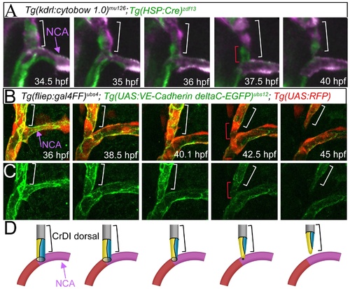
Endothelial cells rearrange from a multicellular into a partially unicellular tube during blood vessel pruning. (A) Still images from a time-lapse movie of Tg(kdrl:cytobow1.0)mu125; Tg(HSP:Cre)zdf13 embryos. Imaged area indicated with box in Figure 1D. Bracket marks dorsal CrDI. At the 37.5 hpf time point, note unicellular connection between the dorsal CrDI and the NCA (single green cell, marked with red bracket). (B) Merged still images from time-lapse imaging of Tg(UAS:RFP); Tg(fliep:Gal4FF)ubs4; Tg(UAS:VE-cadherin-deltaC-EGFP)ubs12 between 36 and 45 hpf. Red bracket at 42.5 hpf indicates unicellular connection between dorsal CrDI and NCA. (C) Still images from Tg(UAS:VE-cadherin-deltaC-EGFP)ubs12 expression corresponding to images in B. (D) Schematic drawing of cell rearrangements shown in B, C. Brackets label dorsal CrDI, purple arrow highlights NCA. |
| Genes: | |
|---|---|
| Fish: | |
| Anatomical Terms: | |
| Stage Range: | Prim-25 to High-pec |