
FIGURE
Fig. 7
- ID
- ZDB-FIG-140122-11
- Publication
- Iwasaki et al., 2013 - Expression of Arginine Vasotocin Receptors in the Developing Zebrafish CNS
- Other Figures
- All Figure Page
- Back to All Figure Page
Fig. 7
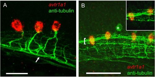
avtr1a1 expressing neurons in the caudal hindbrain project axons into the ipsilateral MLF and are in close proximity with the LLF at 28 hpf. (A) Lateral view (anterior left) showing caudal hindbrain, avtr1a1+ neurons (red) project axons (labeled with anti-acetylated α tubulin) into the ipsilateral MLF (arrow). Scale: 20 µm. (B) Lateral view showing that the LLF (green, bracket) courses over the avtr1a1+ neurons (red). Asterisk denotes a RB sensory neuron. Inset: single confocal plane showing LLF axons coursing over avtr1a1+ hindbrain neurons. Scale: 50 µm. |
Expression Data
| Gene: | |
|---|---|
| Fish: | |
| Anatomical Terms: | |
| Stage: | Prim-5 |
Expression Detail
Antibody Labeling
Phenotype Data
Phenotype Detail
Acknowledgments
This image is the copyrighted work of the attributed author or publisher, and
ZFIN has permission only to display this image to its users.
Additional permissions should be obtained from the applicable author or publisher of the image.
Reprinted from Gene expression patterns : GEP, 13(8), Iwasaki, K., Taguchi, M., Bonkowsky, J.L., and Kuwada, J.Y., Expression of Arginine Vasotocin Receptors in the Developing Zebrafish CNS, 335-42, Copyright (2013) with permission from Elsevier. Full text @ Gene Expr. Patterns