
Fig. 3
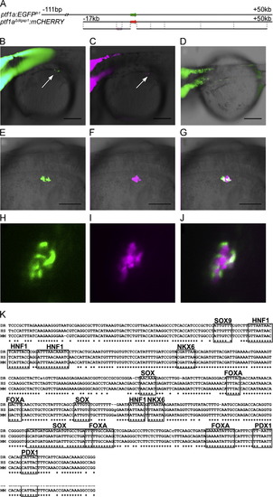
The early pancreatic ptf1a enhancer. (A) The diagram compares the previously published 111 kb to +50 kb ptf1a:egfp BAC transgene, to the truncated -17 kb to +50 kb ptf1aΔdlgap1:mcherry BAC, with only 17 kb of upstream sequence. (B) Expression of egfp from the longer BAC at 34 hpf completely overlaps mCherry expression from the shorter BAC (C), including early expression in the pancreas (arrows in B, C); this early pancreatic expression is not seen with the autoregulatory enhancer (D). (E) The +1ptf1a enhancer is active at 38 hpf in a cluster of cells surrounding the principal islet, marked by expression of the ins:mCherry transgene (F). (H–J) Confocal microscopy of embryos doubly transgenics for +1ptf1a:EGFP (H) and ins:mCherry (I) at 48 hpf show that the two cell populations are adjacent but do not overlap, as seen in a single 5 µM slice (J). (G) A 500 bp core of the +1ptf1a enhancer is highly conserved from mammals to teleosts, and contains predicted binding sites for a number of biologically relevant transcription factors, including Pdx1, Sox9, and Hnf1. DR: zebrafish, HS: human, MM: mouse. Scale bars: 200 μm. |
Reprinted from Developmental Biology, 381(2), Pashos, E., Tae Park, J., Leach, S., and Fisher, S., Distinct enhancers of ptf1a mediate specification and expansion of ventral pancreas in zebrafish, 471-81, Copyright (2013) with permission from Elsevier. Full text @ Dev. Biol.