|
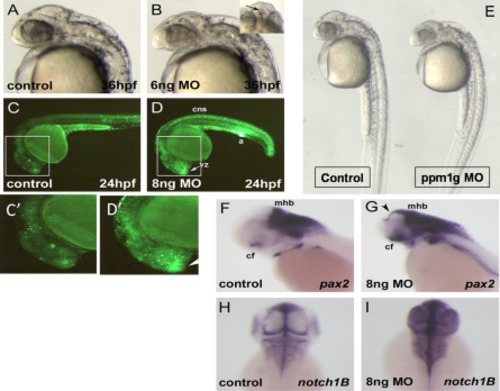
ppm1g morphants have central nervous system defects and aberrant expression of brain markers. A,B: Morphology of embryos injected with 6 ng ppm1gMO or control at 36hpf. Morphants also have necrotic tissue in the presumptive telencephalon (B, inset). C,D: Acridine orange staining indicates elevated apoptosis in the forebrain ventricular zone (vz), central nervous system (cns), and anus (a) in the ppm1g morphants at 24 hpf. C2, D2 are insets enlarged from image areas in C and D as indicated. E: Gross morphology of the whole embryos shown in A and B. In situ hybridization signal for pax2 in ppm1g morphant and control. F,G: Abnormal expression of pax2 in the ppm1g morphant was identified in the midbrain tectum mid and hindbrain, mhb, (arrowhead), choroid fissure (cf), and optic nerve. H,I: Dorsal view of in situ hybridization signal for notch1B in the ppm1gmorphant and control.
|