- Title
-
Nuclear phosphatase PPM1G in cellular survival and neural development
- Authors
- Foster, W.H., Langenbacher, A., Gao, C., Chen, J., and Wang, Y.
- Source
- Full text @ Dev. Dyn.
|
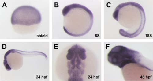
ppm1g expression pattern during zebrafish embryonic development. ppm1g mRNA was detected by in situ hybridization at different developmental stages from the onset of gastrulation (A) to somitogenesis (B,C). Strong expression is shown in the brain, spinal cord, eyes, and branchial arches at both 24 hr post fertilization (hpf) (D, lateral; E, dorsal) and 48 hpf (F). |
|
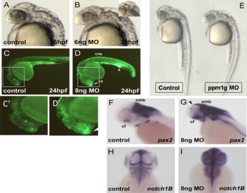
ppm1g morphants have central nervous system defects and aberrant expression of brain markers. A,B: Morphology of embryos injected with 6 ng ppm1gMO or control at 36hpf. Morphants also have necrotic tissue in the presumptive telencephalon (B, inset). C,D: Acridine orange staining indicates elevated apoptosis in the forebrain ventricular zone (vz), central nervous system (cns), and anus (a) in the ppm1g morphants at 24 hpf. C2, D2 are insets enlarged from image areas in C and D as indicated. E: Gross morphology of the whole embryos shown in A and B. In situ hybridization signal for pax2 in ppm1g morphant and control. F,G: Abnormal expression of pax2 in the ppm1g morphant was identified in the midbrain tectum mid and hindbrain, mhb, (arrowhead), choroid fissure (cf), and optic nerve. H,I: Dorsal view of in situ hybridization signal for notch1B in the ppm1gmorphant and control. |
|
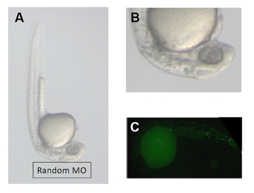
Control morpholino injection for zebrafish development. A. Representative low-amplification image of a whole fish at 32 hpf. B. A high amplification image of brain region from the same fish. C. Acridine orange staining of the whole fish. All of them were injected with a random sequence morpholino (5′-CCT CTT ACC TCA GTT ACA ATT TAT A 3′) at 2 ng. |


