
FIGURE
Fig. 6
- ID
- ZDB-FIG-130906-13
- Publication
- Weger et al., 2013 - Real-time in vivo monitoring of circadian E-box enhancer activity: A robust and sensitive zebrafish reporter line for developmental, chemical and neural biology of the circadian clock
- Other Figures
- All Figure Page
- Back to All Figure Page
Fig. 6
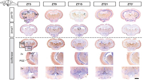
Diurnal time course of per1b, bmal1 and luciferase mRNA expression. Schematic (upper left corner) indicates the level of the sections shown in the figure. Rows: time points at which fish were sacrificed (ZT3, ZT9, ZT15, ZT21, and ZT32 on the following day). Rows show in situ hybridizations with antisense probes for the indicated genes. Rows 4 and 5 show magnifications of the luciferase staining in the optic tectum (TeO, PGZ) and ventral hypothalamus regions (Hv), respectively, as indicated by the boxes in row 3. Scale bar, 250 μm and 125 μm for high magnifications. |
Expression Data
| Genes: | |
|---|---|
| Fish: | |
| Condition: | |
| Anatomical Term: | |
| Stage: | Adult |
Expression Detail
Antibody Labeling
Phenotype Data
Phenotype Detail
Acknowledgments
This image is the copyrighted work of the attributed author or publisher, and
ZFIN has permission only to display this image to its users.
Additional permissions should be obtained from the applicable author or publisher of the image.
Reprinted from Developmental Biology, 380(2), Weger, M., Weger, B.D., Diotel, N., Rastegar, S., Hirota, T., Kay, S.A., Strähle, U., and Dickmeis, T., Real-time in vivo monitoring of circadian E-box enhancer activity: A robust and sensitive zebrafish reporter line for developmental, chemical and neural biology of the circadian clock, 259-73, Copyright (2013) with permission from Elsevier. Full text @ Dev. Biol.