
Fig. S4
- ID
- ZDB-FIG-130828-31
- Publication
- Gupta et al., 2013 - An Injury-Responsive Gata4 Program Shapes the Zebrafish Cardiac Ventricle
- Other Figures
- All Figure Page
- Back to All Figure Page
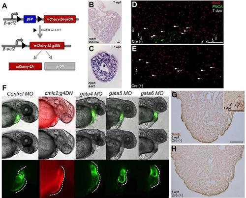
g4DN Overexpression Decreases Cortical Myocyte Proliferation and Phenocopies the gata4 Morphant
(A) Cartoon describing the inducible expression of a dominant-negative version of Gata4. Cre-mediated recombination at loxP sites (black triangles) results in excision of Blue Fluorescent Protein (BFP) and expression of mCherry and g4DN, through a ribosomal 2A based translational skip. (B and C) β-act2:BSg4DN; cmlc2:CreER animals were exposed to vehicle or 4-HT at 30 dpf and raised to 7 wpf. Ventricular sections were then stained by in situ hybridization for nppb; those expressing g4DN had higher cardiac nppb expression (n = 4). (D and E) β-act2:BSg4DN animals with (D) or without (E) the cmlc2:CreER transgene were treated with 4-HT. Three days later, their ventricular apices were resected, and cardiomyocyte proliferation was assessed by Mef2/PCNA staining at 7 dpa. Examples of cortical (black) and trabecular (white) proliferating cardiomyocytes are indicated by arrowheads. (F) Shown (left to right) is a representative control 48 hpf embryo, an embryo transgenic for the cmlc2:g4DN transgene (co-expressing a dominant-negative Gata4 isoform and TagRFP in cardiomycytes), or embryos that had been injected at the one cell stage with morpholinos targeting gata4, gata5, or gata6. Morphant embryonic hearts fluoresce green from a cmlc2:EGFP transgene. Panels show a fluorescent channel (top row), brightfield only (middle row), or a higher magnification view of the heart under fluorescence (bottom row). Dashed lines in the bottom row panels follow the outer curvature of the heart, indicating a normally looped heart tube in the control embryo (25 of 25 injected embryos), a non-looping distended linear heart tubes with chamber demarcation in the cmlc2:g4DN transgenic (28/28) and gata4 morphant (34/36) embryos, a diminutive heart in the gata5 morphant embryo (29/29), and a squat hypoplastic linear heart in the gata6 morphant embryo (27/31). g4DN transgenic embryos from independent founder lines each phenocopied the gata4 morphant, while non-transgenic sibling embryos from these founders developed normal hearts (not shown). (G and H) Apoptosis as assessed by TUNEL staining was not evident after induced cardiomyocyte g4DN expression from 5 to 6 wpf. Representative sections are shown, which indicated only rare TUNEL-positive cardiomyocytes in β-act2:BSg4DN animals with (H, n = 12) or without (G, n = 11) the cmlc2:CreER transgene. Some non-specific edge-staining was present in all samples. Inset shows the resection injury site of a 1 dpa adult ventricle (used as a positive control), indicating several TUNEL-positive cardiomyocytes. Scale bars = 50 μm (B and C); 100 μm (D, E, G, and H). |
| Gene: | |
|---|---|
| Fish: | |
| Knockdown Reagents: | |
| Anatomical Term: | |
| Stage: | Long-pec |
| Fish: | |
|---|---|
| Knockdown Reagents: | |
| Observed In: | |
| Stage: | Long-pec |