
Fig. 3
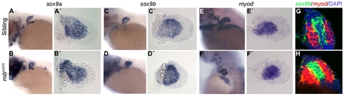
Cartilage and muscle masses are specified in mibta52b mutants. Whole-mount in situ hybridization of 68 hpf siblings (n = 15) (A, A′), (n = 12) (C, C′), (n = 14) (E, E′) and mibta52b mutants (n = 12) (B, B′), (n = 15) (D, D′), (n = 11) (F, F′) using sox9a and sox9b as cartilage markers and myod as a muscle marker, showing a slight upregulation of these genes in mibta52b mutant pectoral fin. (A-F) Whole larvae. (A′-F′) Detached pectoral fins with distal to the right. Double fluorescent in situ hybridization on sections using sox9b (green) and myod (red), counterstained with DAPI to label the nuclei (blue) shows no signs of cell mixing in a sibling (n = 7) (G) and a mibta52b mutant pectoral fins (n = 9) (H). An upregulation of these genes is observed in mibta52b mutant pectoral fins when compared with the sibling. |
| Genes: | |
|---|---|
| Fish: | |
| Anatomical Terms: | |
| Stage: | Pec-fin |
| Fish: | |
|---|---|
| Observed In: | |
| Stage: | Pec-fin |