
Fig. 8
- ID
- ZDB-FIG-130719-8
- Publication
- Riera et al., 2013 - CERKL Knockdown Causes Retinal Degeneration in Zebrafish
- Other Figures
- All Figure Page
- Back to All Figure Page
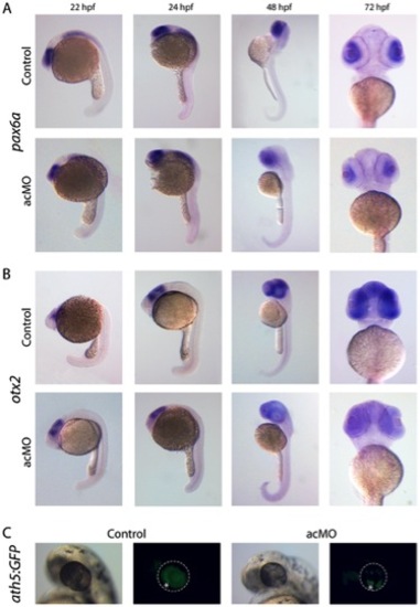
Expression of retina cell markers in acMO-injected embryos at early developmental stages. (A and B) At 22 and 24 hpf, the spatiotemporal pattern of pax6a (A) and otx2 (B) in acMO-injected animals was similar to that of controls: pax6a was detected in the forebrain, hindbrain, spinal cord and eye, and otx2 in the eye and midbrain. By 48 and becoming more evident at 72 hpf, acMO embryos exhibited a marked reduction in the expression of both markers. (C) The expression of the ath5 transcription factor was assessed in vivo in acMO-injected embryos of the transgenic ath5:GFP strain. At 48 hpf, the wave of ath5 expression, which prefigures the wave of retinal ganglion cell genesis, filled the central and peripheral retina of control embryos, whereas the pattern appeared delayed and disorganized in the acMO morphants, although RGC genesis was not fully abolished. * denotes the ventronasal patch of RGC genesis. |
| Genes: | |
|---|---|
| Fish: | |
| Knockdown Reagent: | |
| Anatomical Terms: | |
| Stage Range: | 26+ somites to Protruding-mouth |