
Fig. 5
- ID
- ZDB-FIG-130429-16
- Publication
- Liu et al., 2013 - Direct and indirect roles of Fgf3 and Fgf10 in innervation and vascularisation of the vertebrate hypothalamic neurohypophysis
- Other Figures
- All Figure Page
- Back to All Figure Page
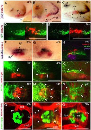
fgf3 expression and NH innervation and vascularisation in zebrafish. Lateral (A-K) or ventral (L-Q) views of wild-type zebrafish; anterior to the left. (A-C) fgf3 expression in posterior cells of the crabp1a+ NH (overlap in A indicated by arrow) and ventral-most cells of the posterior hypothalamus from 33 to 72 hpf. Dotted outline in B,C shows AH. (D,E) Double fluorescent in situ hybridisation reveals a partial overlap of fgf3 (green) and crapb1a (red) expression in posterior regions of the crabp1a domain at 32 hpf (D), whereas at 36 hpf, fgf3+ cells lie exclusively posterior of the crabp1a domain (E). (F,G) The crabp1a+ NH lies directly dorsal to the pomca+ AH. (H) Triple staining reveals innervation (AcTub) of NH (crabp1a) directly dorsal to AH (prl:RFP transgene product in lactotropes). Note that expression of AcTub and crabp1a signals are suboptimal owing to different fixation requirements. (I-N) Pioneering axons enter the anterior/dorsal NH anlage at 36 hpf (I,L). Axons are detected in the anterior half of the NH at 48 hpf (J,M), and throughout the NH at 72 hpf (K,N). Axons within the NH are indicated by arrows, axons outside the NH with arrowheads. For spatial reference, specimens are counterstained for the AH marker pomca. (O-Q) Endothelial cells (arrowhead) are detected at 36 hpf close to anterior regions of the pituitary (marked by pomca expression in AH) (O). At 48 hpf, endothelial cells assembling to vessels are detected anteriorly (arrowheads) and posteriorly (arrows) (P). At 72 hpf, the hypophyseal blood vessel system is completed (Q). ah, adenohypophysis; bca, basal communicating artery; cadi, caudal division of internal carotid artery; h, hours post fertilisation (hpf); hya, hypophyseal artery; hyc; hypophyseal capillary; hyv, hypophyseal vein; nh, neurohypophysis; oc, oral cavity; os, optic stalk. Scale bars: 50 μm. |