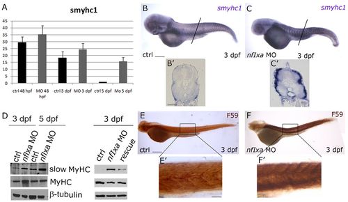
Slow myosin remains expressed in nfixa-MO injected embryos after 48 hpf. (A) Quantitative real-time PCR (qRT-PCR) of smyhc1 mRNA expression normalized to ef1a. The expression of smyhc1 in control embryos presents a peak at 48 hpf and is then downregulated at 3 and 5 dpf. In nfixa-MO-injected embryos, the expression of smyhc1 remains elevated even after 48 hpf. (B-C′) Whole-mount in situ hybridization with smyhc1 probe replicates qRT-PCR results: at 3 dpf, nfixa-MO-injected embryos (C,C′) still present a stronger signal of smyhc1 than controls (B,B′). B′ and C′ are transverse sections of the tail. (D) Western blot: slow myosins are recognized by F59 antibody; all sarcomeric myosins are recognized by MF20 antibody. At 3 and 5 dpf in nfixa-MO injected embryos, slow and all myosin protein levels are higher than the control, whereas protein levels were recovered in rescued embryos at 3 dpf. Additional lower molecular weight bands in the 3 dpf nfixa-MO MyHC lane correspond to degradation products of the protein. (E-F′) Immunostaining with F59 antibody (slow fibers): at 3 dpf, nfixa-MO-injected embryos (F,F′) present a stronger F59 signal than the control (E,E′). Insets in E2 and F2 show a higher magnification of the tail region. In all figures, lateral views are shown, anterior is always towards the left. Scale bars: 100 μm in B,B′,C,C′,E,F; 200 μm in E′,F′.
|
