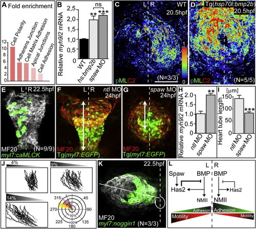
Control of Cardiac Progenitor Cell Motility by Bmp(A) Enrichment analysis of genes upregulated in Tg[hsp70l:bmp2b]fr13 versus WT cardiac tissue at 21.5 hpf according to Database for Annotation, Visualization and Integrated Discovery-based clustering for functional terms.(B) RT-qPCR comparative analysis of relative expression levels of myh9l2 in cardiac tissue-derived cDNAs at 24 hpf (mean values with SEM; p < 0.01; p < 0.0005).(C) Expression of phosphorylated NMII (pMLC2) is slightly higher on the right side of the WT cardiac field.(D) Strong expression of phosphorylated NMII throughout the entire heart cone in Tg[hsp70l:bmp2b]fr13 transgenic embryos heat shocked at 18 hpf.(E) Clonal misexpression of caMLCK on the left side of the cardiac cone affects cardiac laterality.(F and G) Loss of Ntl (F) and Spaw (G) abolishes cardiac laterality, but heart tube elongation is most strongly affected in spaw morphants. Heart tube length is the extension between the outflow tract region and leading edge of the atrium (arrows).(H) RT-qPCR comparative analysis of relative expression levels of myh9l2 in spaw and ntl morphant hearts at 24 hpf (mean values with SEM; p < 0.005).(I) Heart tube length in spaw and ntl morphants at 24 hpf (mean values with SEM; p < 0.0005).(J) Simulations of cardiac jogging based on a random cell motility model. Three representative simulations covering a range of L/R differences in cell motility and the respective bearing angles based on ten independent simulations for each condition are shown. The 9% condition of L/R motility differences most closely resembles WT cardiac jogging (red bearing angle).(K) Clonal misexpression of the Bmp antagonist Noggin1 within the left myocardium enhances the angle of cardiac jogging toward the left.(L) Model of Nodal-Bmp pathway interactions during stages of cardiac jogging.Total numbers of embryos indicate the occurrence of the most common phenotypes as shown. L, left; R, right; ns, not significant (for details on statistical analysis, see also Supplemental Experimental Procedures). Scale bars, 50 μm.See also Figures S3 and S4.
|
