
Fig. 2
- ID
- ZDB-FIG-130326-29
- Publication
- Ravi et al., 2013 - Sequencing of pax6 Loci from the elephant shark reveals a family of pax6 genes in vertebrate genomes, forged by ancient duplications and divergences
- Other Figures
- All Figure Page
- Back to All Figure Page
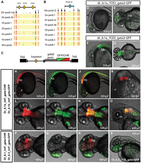
Comparison of functional activity driven by zebrafish and medaka intron 7 sequences. A, B) Comparison of full Pax6 intron 7 sequences of multiple teleost species visualized as PIP plots. A) PIP plot with zebrafish Pax6.1a intron 7 as baseline sequence against intron 7 sequences of the Pax6.1 and Pax6.3 genes of zebrafish (Danio rerio (Dr)), medaka (Oryzias latipes (Ol)), stickleback (Gasteroteus aculeatus (Ga)), tetraodon (Tetraodon nigroviridis (Tn)) and mouse (Mus musculus (Mm)), showing lack of sequence conservation of Pax6.1 intron 7 with the Pax6.3 loci. A variable subset of CNEs has been conserved in the Pax6.1 loci. B) PIP plot using stickleback intron 7 as base sequence against the intron 7 regions of the Pax6.1 and Pax6.3 loci from multiple teleosts, showing the presence of a conserved element unique to the Pax6.3 genes only. C) To assess whether patterns of sequence conservation are reflected at the functional cis-regulatory level the full intron 7 sequences from zebrafish Pax6.1a (Dr6.1a_int7), zebrafish Pax6.1b (Dr6.1b_int7), medaka Pax6.1 (Ol6.1_int7) and medaka Pax6.3 (Ol6.3_int7) were cloned in front of a gata2 minimal promoter-reporter cassette in a Tol2-2way reporter vector system. Most fragments were cloned with both GFP and mCherry as fluorescent reporter to allow combinatorial analysis in dual fluorescence reporter transgenic zebrafish. (D–G) In addition smaller fragments containing the individual 7CE1, 7CE2 and 7CE3 elements from the zebrafish Pax6.1a gene, and the 7CE(6.3) element from the medaka Pax6.3 gene were also cloned and used to produce transient transgenic zebrafish. (D, E) Lateral and dorsal views of transgenic fish for the Dr6.1a_7CE2 element show expression in the diencephalon (d) at 30 and 48 hours post fertilization (hpf). (F, G) Lateral and dorsal views of transgenic fish for the 7CE3 element of zebrafish Pax6.1a show expression in the hindbrain (hb) at 30 and 48 hpf. (H–O) Expression driven by the full intron 7 sequences of zebrafish Pax6.1a and Pax6.1b is consistent with the presence or absence of the 7CE2 and 7CE3 elements. mCherry fluorescence at 30 and 48 hpf recapitulates the combined pattern of the 7CE2 and 7CE3 elements with expression seen in hindbrain (hb) and diencephalon (d). GFP fluorescence, driven by zebrafish Pax6.1b intron 7 is observed in the hindbrain and neural tube of transgenic fish, but no signal is seen in the diencephalon in accordance with the absence of the 7CE2 element from the Dr6.1b intron7. A difference in the detail of hindbrain/neural tube expression driven by the Dr6.1a and 6.1b intron 7 sequences is also observed with a stronger and wider expression of Dr6.1a_int7 in the hindbrain and decreasing towards the caudal neural tube, while Dr6.1b_int7 driven expression is narrower in the hindbrain but maintained more evenly along the neural tube. l, lens; nr, neuroretina. (P–S) Medaka Pax6.1 intron 7 drives expression in a similar pattern to zebrafish Pax6.1a with clear expression in diencephalon and hindbrain, with decreasing levels in the neural tube, in accordance with the conservation of the 7CE2 and 7CE3 elements in the intron. Medaka Pax6.3 intron 7 (Ol6.3_int7) drives GFP reporter expression in the eye (e) of transgenic zebrafish, and this expression is replicated when the 7CE(6.3) element, conserved only in Pax6.3 loci, is used on its own. |
| Genes: | |
|---|---|
| Fish: | |
| Anatomical Terms: | |
| Stage Range: | Prim-15 to Long-pec |