
Fig. 11
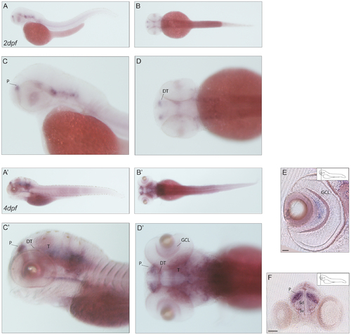
Expression pattern of caln2. (A–D2) mRNA expression of caln2 in lateral (A,C–A2,C2) and dorsal (B,D–B2D2) views of 2dpf (A–D) and 4dpf (A2–D2) larvae with zoom in (C,D–C2,D2). The onset of caln1 expression takes place within the first two days of embryonic development. (A–D) At 2dpf the caln1 transcript localizes to the forebrain at the level of the subpallial region and in the caudal midbrain. (A-‘D’) The same expression profile seems to be maintained until day 4 of development. (E) Transverse section of the retina showing signal of caln1 antisense probe in a subset of retinal ganglion cells in the central part of the ganglion cell layer. Strongly stained cells are present in the region of the telencephalic pallial domain (F). Scale bar: 50 µm. P: pallium; DT: dorsal thalamus; T: midbrain tegmentum; GCL: ganglion cell layer; Sd: dorsal division of subpallium; Sv: ventral division of subpallium. |
| Gene: | |
|---|---|
| Fish: | |
| Anatomical Terms: | |
| Stage Range: | Long-pec to Day 4 |