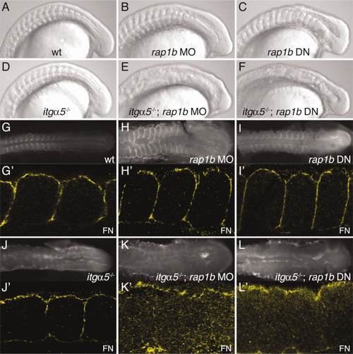
Loss of rap1b synergizes with itgα5, resulting in a disruption of all somite borders and abrogation of Fibronectin matrix assembly. A–F: Lateral view DIC images showing clear somite borders for (A) wild-type, (B) rap1b MO1-injected (8 experiments, 83.7% affected, n = 341), and (C) rap1b DN mRNA-injected (3 experiments, 100% affected, n = 177) embryos. For synergy experiments, rap1b MO1 or rap1b DN was injected into the progeny of heterozygous itgα5+/- crosses. Therefore, 100% penetrance would result in approximately 25% of embryos exhibiting the phenotype. D: Anterior somite borders are disrupted in the itgα5-/- mutant, while (E) itgα5-/-; rap1b MO1 (9 experiments, 27.4% affected, n = 865) and (F) itgα5-/-; rap1b DN (6 experiments, 22.3% affected, n = 455) embryos lack somite borders along the entire AP axis. (G- L) Dorsal view and (G′-L′) higher magnification dorsal view of FN localization showing clear FN matrix surrounding somites in (G–G′) wild-type, (H–H′) rap1b MO1, and (I–I′) rap1b DN embryos. J,J′: FN matrix surrounds only posterior somites in itgα5-/- mutants, but few fibrils are seen within the paraxial mesoderm of either (K,K′) itgα5-/-; rap1b MO1 or (L,L′) itgα5-/-; rap1b DN embryos. For all images, anterior is to the left and embryos are at the 12–15-somite stage.
|
