
Fig. S1
- ID
- ZDB-FIG-130118-7
- Publication
- Freitas et al., 2012 - Hoxd13 contribution to the evolution of vertebrate appendages
- Other Figures
- All Figure Page
- Back to All Figure Page
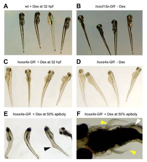
Control experiments for the hoxd13a and hoxa4a dexamethasone (Dex)-inducible injected constructs. All panels show embryos at 4 dpf. A-B, Wild type embryos (wt) treated with Dex 32 hpf (A) and embryos injected with hoxd13a-GR but not treated with Dex (B) did not show any phenotypic alteration. C-D, Control embryos injected with hoxa4a-GR, did not present phenotypic alterations either when they were exposed (C) or not (D) to Dex at 32 hpf. E-F, Deformation of the body axis was observed in 18% (n=50) of sibling embryos injected with hoxa4a-GR and treated with Dex at 50% of epiboly (E) but no abnormalities were detected in fins (yellow arrows, F). |
Reprinted from Developmental Cell, 23(6), Freitas, R., Gómez-Marín, C., Wilson, J.M., Casares, F., and Gómez-Skarmeta, J.L., Hoxd13 contribution to the evolution of vertebrate appendages, 1219-1229, Copyright (2012) with permission from Elsevier. Full text @ Dev. Cell