
FIGURE
Fig. 5
- ID
- ZDB-FIG-130108-5
- Publication
- Uribe et al., 2012 - Id2a functions to limit Notch pathway activity and thereby influence the transition from proliferation to differentiation of retinoblasts during zebrafish retinogenesis
- Other Figures
- All Figure Page
- Back to All Figure Page
Fig. 5
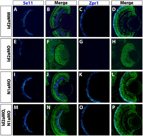
Knockdown of Notch1a rescues terminal differentiation of retinal neurons in Id2a-deficient retinae. Transverse retinal sections from Id2aMM (A)–(D), Id2aMO (E)–(H), N1aMO (I)–(L) and (M)–(P) Id2aMO/N1aMO embryos at 72 hpf assayed for 5e11 expression (amacrine cells) or Zpr1 expression (red/green cones). Merged images show co-staining of retinal marker (blue) and nuclei (Sytox-green; green). Dorsal is up in all panels. |
Expression Data
Expression Detail
Antibody Labeling
Phenotype Data
| Fish: | |
|---|---|
| Knockdown Reagents: | |
| Observed In: | |
| Stage: | Protruding-mouth |
Phenotype Detail
Acknowledgments
This image is the copyrighted work of the attributed author or publisher, and
ZFIN has permission only to display this image to its users.
Additional permissions should be obtained from the applicable author or publisher of the image.
Reprinted from Developmental Biology, 371(2), Uribe, R.A., Kwon, T., Marcotte, E.M., and Gross, J.M., Id2a functions to limit Notch pathway activity and thereby influence the transition from proliferation to differentiation of retinoblasts during zebrafish retinogenesis, 280-292, Copyright (2012) with permission from Elsevier. Full text @ Dev. Biol.