
FIGURE
Fig. 4
- ID
- ZDB-FIG-130108-4
- Publication
- Uribe et al., 2012 - Id2a functions to limit Notch pathway activity and thereby influence the transition from proliferation to differentiation of retinoblasts during zebrafish retinogenesis
- Other Figures
- All Figure Page
- Back to All Figure Page
Fig. 4
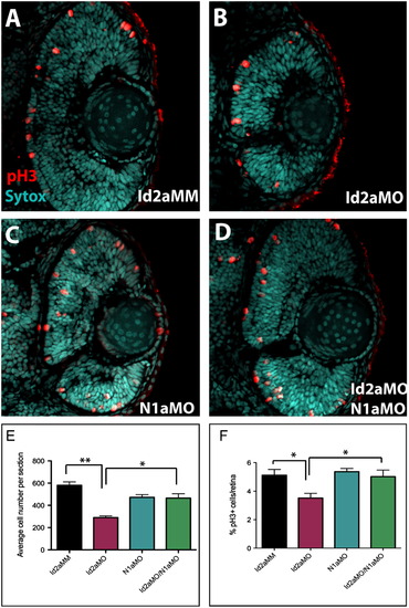
Knockdown of Notch1a rescues retinoblast proliferation defects in Id2a-deficient retinae. (A)–(D) pH3 immunohistochemistry was used to quantify retinoblasts in late G2/M in transverse retinal sections from (A) Id2aMM, (B) Id2aMO, (C) N1aMO and (D) Id2aMO/N1aMO embryos at 48 hpf. Average total cell number per retinal section was determined for each condition (E), as well as the average percentage of pH3-positive cells per retinal section (F). Dorsal is up in all images. Error bars represent SEM, n=9; *p<0.05, ** p<0.005. |
Expression Data
Expression Detail
Antibody Labeling
Phenotype Data
| Fish: | |
|---|---|
| Knockdown Reagents: | |
| Observed In: | |
| Stage: | Long-pec |
Phenotype Detail
Acknowledgments
This image is the copyrighted work of the attributed author or publisher, and
ZFIN has permission only to display this image to its users.
Additional permissions should be obtained from the applicable author or publisher of the image.
Reprinted from Developmental Biology, 371(2), Uribe, R.A., Kwon, T., Marcotte, E.M., and Gross, J.M., Id2a functions to limit Notch pathway activity and thereby influence the transition from proliferation to differentiation of retinoblasts during zebrafish retinogenesis, 280-292, Copyright (2012) with permission from Elsevier. Full text @ Dev. Biol.