Fig. 6
- ID
- ZDB-FIG-130102-6
- Publication
- Vemaraju et al., 2012 - A spatial and temporal gradient of fgf differentially regulates distinct stages of neural development in the zebrafish inner ear
- Other Figures
- All Figure Page
- Back to All Figure Page
|
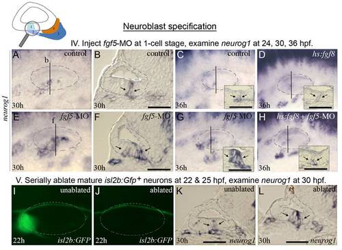
fgf5 from mature neurons terminates the phase of neuroblast specification. The icon at the top of the figure indicates that analysis focuses on initial formation of neuroblasts. Experimental manipulations in groups IV and V are briefly summarized at the tops of the corresponding data panels. (A-H) Expression of neurog1 in control embryos (A-C), a hs:fgf8 embryo (D), fgf5 morphants (E-G), and a hs:fgf8 embryo injected with fgf5-MO (H) at the indicated stages. Transgenic embryos (D, H) were heat shocked for 30 minutes at 39°C beginning at 24 hpf. Vertical lines in (A, C-E, G, H) indicate the plane of transverse sections in (B, F, and insets in C, D, G and H). (I-L) Expression of isl2b:Gfp at 22 hpf (I, J) and neurog1 at 30 hpf (K, L) in a specimen in which mature (fgf5-expressing) neurons were laser-ablated. The same specimen is shown in all panels. Mature SAG neurons expressing isl2b:Gfp were serially ablated on the left side at 22 hpf (J) and 25 hpf (not shown), and the embryo was fixed and sectioned at 30 hpf to examine neurog1 expression (L). Images of the unablated right side (I, K) were inverted to facilitate comparison. The surface of the otic vesicle is outlined in all panels. Arrows in sections indicate the edges of neurog1 domain in the otic floor. Note that the amount and duration of delamination of neurog1+ neuroblasts is strongly enhanced by knockdown of fgf5 (F, G) or ablation of mature neurons (L). Activation of hs:fgf8 reverses the effects of fgf5-MO (H). Scale bar, 25 μm. Transverse sections are shown with lateral to the left and dorsal up. Wholemount images show dorsolateral views with anterior to the left. |
| Genes: | |
|---|---|
| Fish: | |
| Condition: | |
| Knockdown Reagent: | |
| Anatomical Terms: | |
| Stage Range: | 26+ somites to Prim-25 |

