
Fig. 3
- ID
- ZDB-FIG-121102-2
- Publication
- Ichimura et al., 2012 - A comparative analysis of glomerulus development in the pronephros of medaka and zebrafish
- Other Figures
- All Figure Page
- Back to All Figure Page
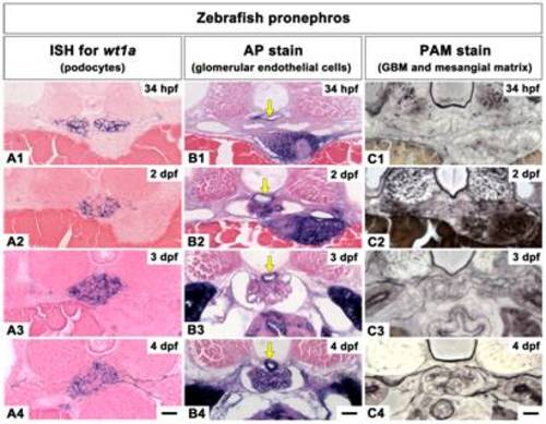
Temporal specification of the three glomerular components during glomerular development in zebrafish. A1-A4: wt1a mRNA is expressed in the forming glomerulus from 34 hpf to 4 dpf. Cross sections show wt1a-positive podocyte layers of the paired glomeruli at 34 hpf (A1). Unlike medaka, zebrafish wt1a-positive podocytes are merged at 2 dpf (A2). B1-B4: Glomerular capillaries are detected by AP stain. At 34 hpf, the flattened nephron primordia show proximity to the dorsal aorta (B1). Glomerular capillaries are found at 2 dpf (B2). Dorsal aorta (arrows in B1-B4). C1-C4: GBM and mesangial matrix are detected by PAM stain. The paired glomeruli fuse at the midline and the interglomerular mesangium is not formed (C2-C4). Scale bars = 10 μm. |