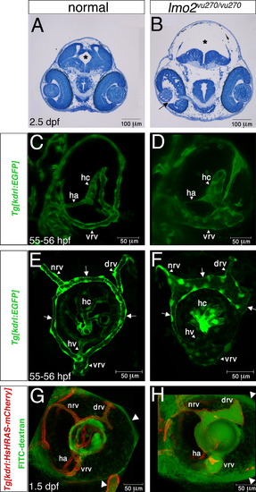
Edema and ocular vessel phenotypes in lmo2 mutant embryos. (A and B) Transverse sections of heads from normal (A) and lmo2 mutant (B) embryos at 2.5 dpf. In the mutant note enlarged ventricle and white spaces within head and eye (arrow) tissues suggesting severe edema. Asterisks mark brain ventricle. (C and D) Three-dimensional shadow rendering confocal images of 55–56 hpf Tg(kdrl:EGFP) normal (C) and lmo2 mutant (D) eyes in frontal view. (E and F) Three-dimensional maximum projection confocal images of 55–56 hpf Tg(kdrl:EGFP) normal (E) and lmo2 mutant (F) eyes in lateral view. Arrows point at different regions of the superficial ring vessel. (G and H) FITC-dextran microangiography of Tg(kdrl:HsHRAS-mCherry) normal (G) and lmo2 mutant (H) eyes in live embryos at 1.5 dpf, lateral view. Note accumulation of dye around the mutant eye (arrowheads). drv, dorsal radial vessel; ha, hyaloid artery; hc, hyaloid capillaries; hv, hyaloid vein; nrv, nasal radial vessel; vrv, ventral radial vessel. In (E–H) anterior is to the left.
|
