
Fig. 3
- ID
- ZDB-FIG-120824-30
- Publication
- Won et al., 2012 - Characterization of na(+) and ca(2+) channels in zebrafish dorsal root ganglion neurons
- Other Figures
- All Figure Page
- Back to All Figure Page
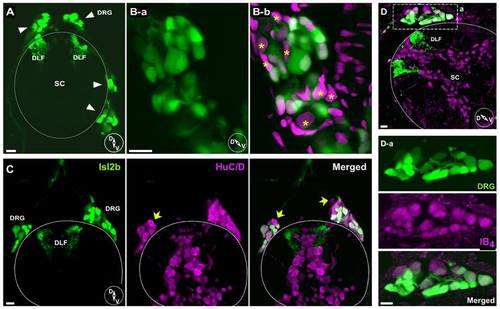
Immunohistochemistry of DRG neurons in juvenile isl2b:EGFP fish sections. A, Confocal image displaying EGFP-labeled DRG neurons and DFL in transverse section. Note that both dorsal and ventrolateral positioned DRG (arrows) cell bodies were observed. B, Higher magnification images of DRG ganglion reveal a variety of cell body sizes (B-a). Counterstaining with DAPI (B-b, magenta) revealed that some EGFP-negative or dim cell bodies were also present in DRG sections (asterisks). C, EGFP-positive and -negative DRG neurons from juvenile isl2b:EGFP fish. A few DRG neurons stained with anti-HuC/HuD neuronal protein antibody (magenta) did not express EGFP. D, Isolectin B4 (IB4) staining. Most DRG cell bodies and some spinal neurons were labeled with IB4 (magenta). Dashed rectangular region was enlarged for displaying labeled DRG neurons (D-a). Solid line represents the dorsal boundary of the spinal cord. Inset cartoons represent orientation of images. D, dorsal; V, ventral; SC, spinal cord; DFL, dorsal longitudinal fasciculus. All scale bars represent 10 μm. |