|
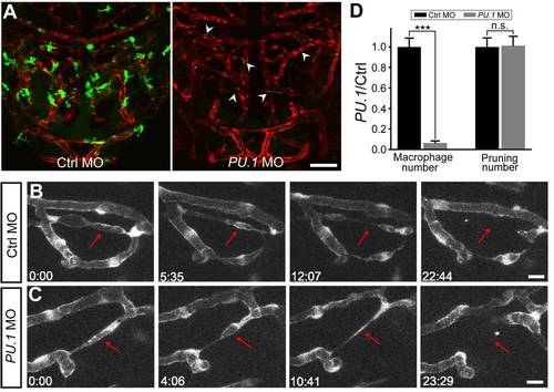
Macrophages are not required for vessel pruning. (A) Projected confocal images showing midbrain vasculature (red) and macrophages (green) in control MO- (left) and PU.1 MO-injected (right) Tg(kdrl:RFP,PU.1:gal4-uas-GFP) larvae at 3 dpf. The green signals in the vessels (arrowheads) were originated from non-specific expression of GFP in blood cells. (B and C) Representative of serial images showing vessel pruning (arrows) in a control MO- (B) and PU.1 MO-injected (C) larvae. (D) Summary of PU.1 knockdown effects on macrophage development and vessel pruning occurrence in the zebrafish midbrain. The data were obtained from 16 larvae in each group. Scales, 50 μm in (A) and 10 μm in (B). n.s., no significance; *** p<0.001 (Student′s t test). Error bars, ± SEM.
|