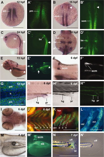
Comparison of aldh1a2 gene expression and aldh1a2:gfp reporter fluorescence. Whole mount in situ hybridization patterns of aldh1a2 gene expression resemble those of aldh1a2:gfp fluorescence. Shifting patterns of gene expression can be observed in the paraxial mesoderm (pm) (A, A′), developing somites (som) and head mesoderm (arrowhead, B, B′), dorsal retina (dr) and cells enclosing the anterior spinal cord at the level of the first 2 somites (arrow) (C, C′), branchial arch (ba) primordia, pectoral fin mesenchyme (pf) and head mesenchyme (hm) contributing to the otic region (D, D′). After hatching, additional expression domains are visible in the branchial arches (arrow), pronephric tubules (arrowhead), and spinal cord motorneurons (scm) at pectoral fin level (E–F′). Higher magnification of paraxial mesodermal cells at 12 hpf shows cytoplasmic aldh1a2 expression; notochordal cells (n) are unlabeled (G). At 6 dpf, expression is apparent in the intestinal walls of the hindgut (hg) (H–H′′), as well as in cells located distal and posterior to the first 4 branchial arches (pharyngeal arches 3–6), which are gill-bearing arches (I–L). GFP fluorescence (green channel) is distinct from chondrocytes (marked in red by a col2a1:mCherry transgenic line). Nuclei were counterstained with DAPI in embryos in G, L. The swim bladder (sb) expresses aldh1a2 at 4 dpf (M, M′). aldh1a2 is upregulated in the regenerating caudal fin fold (cff), 2 days after amputation (N), whereas unamputated controls show no reporter expression (N′). All images are oriented with rostral to the left; A–B′, D, D′, G: dorsal views; C, C′, E–F′, H, H′, K, M–N′: lateral views; I, J, L: ventral views. F′, G, H′, L: aldh1a2:gfp labeled with anti-GFP antibody. Scale bars = 250 μm (A–F, I, M, N), 100 μm (J, K), 50 μm (G, H, L). bl, blastema-like cells; cb, ceratobranchial cartilage; ch, ceratohyal cartilage; hyo, hyosymplectic cartilage; pr, proctodeum/cloaca.
|
