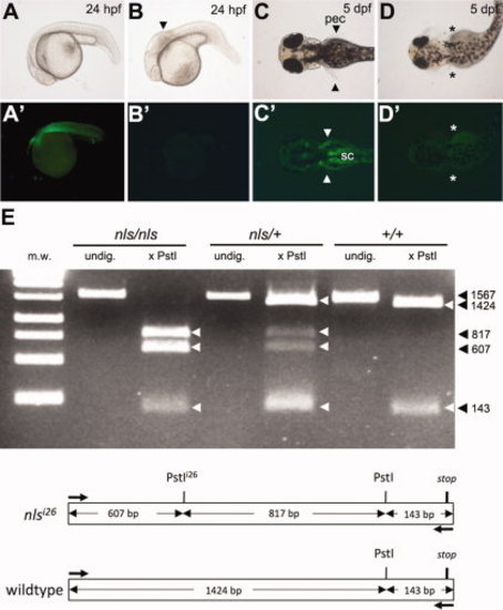
The aldh1a2:gfp transgene rescues the neckless (nls) mutant phenotype. A–D′: Offspring from an incross between nls -/-; aldh1a2-GFP +/- fish (see Table 1). All GFP-positive embryos have wild type appearance (A,C,), whereas all GFP-negative embryos (B′, D′) show the nls mutant phenotype (B,D), including a shortened hindbrain with kinked appearance (arrowhead, B) and absence of pectoral fins (asterisks, D). GFP expression in pectoral fins (pec, arrowheads) and spinal cord (sc) motorneurons at pectoral level (C′) contrasts with autofluorescence of the yolk in all embryos at 5 days post-fertilization (dpf) (C′,D′). E: PstI-digestion of a 1,567-bp endogenous aldh1a2 PCR fragment amplified from cDNA detects a restriction fragment polymorphism in the presence of the mutant nlsi26 allele. cDNA was prepared from regenerating caudal fin blastema of adult aldh1a2-GFP fish predicted to be homozygous, heterozygous, or wt for the nlsi26 allele (Table 1). Schematic shows restriction map of the amplified region. m.w., molecular weight ladder; undig., undigested.
|
