
Fig. 5
- ID
- ZDB-FIG-120525-20
- Publication
- Telfer et al., 2012 - neb: a zebrafish model of nemaline myopathy due to nebulin mutation
- Other Figures
- All Figure Page
- Back to All Figure Page
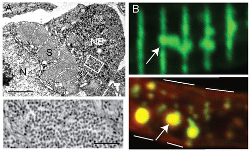
Nemaline bodies in neb skeletal muscle. (A) Ultrastructural analysis reveals the presence of aggregates similar to nemaline bodies. Top panel: a typical example of a nemaline body (NB) is depicted in a myofiber examined in cross-section. S, sarcomere; N, nucleus. Scale bar: 1 μm. Bottom panel: higher magnification of the area denoted by the white box demonstrates that the aggregates are composed primarily of filamentous material. Scale bar: 250 nm. (B) Immunohistochemical analysis of isolated myofibers from 3-dpf neb embryos using an antibody to α-actinin. Top panel: frequent areas of aberrant α-actinin accumulation were detected (arrow), consistent with the presence of nemaline bodies. Bottom panel: co-staining with phalloidin (red) reveals the presence of actin in the aggregated areas (arrow). White lines denote the sarcolemmal membrane. |
| Fish: | |
|---|---|
| Observed In: | |
| Stage: | Protruding-mouth |