
Fig. S3
- ID
- ZDB-FIG-120330-17
- Publication
- Theunissen et al., 2011 - Reprogramming capacity of Nanog is functionally conserved in vertebrates and resides in a unique homeodomain
- Other Figures
- All Figure Page
- Back to All Figure Page
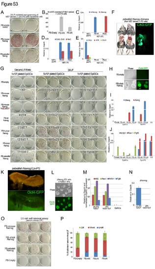
Vertebrate orthologs of Nanog induce naive pluripotency in multiple cell systems. (A) AP staining of Nanog-/- MEF-derived pre-iPS cells transfected with PB transgenes containing an empty vector, mNanog or zNanog, and subsequently cultured in 2i/LIF conditions for 10 days. (B) Quantification of the total number of AP-positive iPS cell colonies at day 10 after medium switch. Error bars indicate one s.d. (n=2). (C) qRT-PCR analysis confirming the presence of mNanog or zNanog transcript in MEF-derived iPS-/- cells generated with mNanog or zNanog. Error bars indicate 1s.d. (D) qRT-PCR analysis for endogenous pluripotency gene expression in MEF-derived iPS-/- cells generated with mNanog or zNanog. Error bars indicate 1 s.d. (E) qRT-PCR analysis for retroviral transgene expression in MEF-derived iPS-/- cells generated with mNanog or zNanog. Error bars indicate 1 s.d. (F) Adult chimeric mouse obtained after blastocyst injection of MEF-derived zNanog iPS-/- cells following Cre excision of the loxP-flanked Nanog transgene. GFP indicates iPS cell contribution. (G) AP staining of EpiSCs transfected with PB transgenes containing an empty vector, mNanog, mNanogY42E, K43T, rNanog, hNanog, cNanog or zNanog, and subsequently cultured for 10 days in presence of the MEK inhibitor PD0325901 and LIF or 2i/LIF. Average numbers of colonies in each condition are indicated. Error bars indicate 1 s.d. (H) Phase and GFP images of emerging iPS cell colonies during 2i/LIF induction of EpiSCs transfected with an empty vector or a cNanog PB transgene. Fluorescence indicates activation of the Oct4-GFP-ires-puro reporter transgene. (I) qRT-PCR analysis confirming the presence of zNanog or cNanog transcript in EpiSC-derived iPS cells generated with cNanog or zNanog. Error bars indicate 1 s.d. (J) qRT-PCR analysis of Nr0b1,Rex1 and Fgf5 expression in EpiSC-derived iPS cells generated with zNanog or cNanog. Error bars indicate 1 s.d. (K) Genital ridge from chimeric embryo dissected at E12.5 shows contribution of EpiSC-derived iPS cells generated with zNanog, which express an Oct4-GFP transgene, to the germline. (L) Phase and GFP images of iPS cells generated from EpiSCs with zNanog in serum-free medium with LIF, and subsequently passaged for 10 passages in serum-free medium alone. Fluorescence indicates activation of the Oct4-GFP-ires-puro reporter transgene. (M) qRT-PCR analysis of Klf2, Rex1, Nr0b1 and Fgf4 expression in iPS cells generated with zNanog in serum-free medium with LIF, and subsequently passaged for 10 passages in serum-free medium alone. Error bars indicate 1 s.d. (N) qRT-PCR analysis confirming zNanog expression in iPS cells generated with zNanog in serum-free medium with LIF, and subsequently passaged for 10 passages in serum-free medium alone. Error bars indicate 1 s.d. (O) AP staining of ES cells transfected with mNanog, cNanog and zNanog, and plated at clonal density in serum minus LIF. (P) Quantification of the proportion of undifferentiated, mixed and differentiated colonies in each condition shown in O. Error bars indicate 1 s.d. (n=3). Representative examples of colonies in each category are shown in Fig. 3M. |