
Fig. 4
- ID
- ZDB-FIG-120329-48
- Publication
- Royo et al., 2012 - Identification and Analysis of Conserved cis-Regulatory Regions of the MEIS1 Gene
- Other Figures
- All Figure Page
- Back to All Figure Page
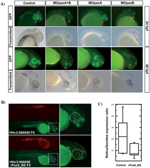
Pax6 regulates the enhancer activity of HHc2:066650. A) Representative pictures of transgenic zebrafish embryos at 24 and 48 hpf, corresponding to HHc2:066650 F2 offspring injected with MOpaxA, MOpaxB, or a mixture of both. The red arrow points to the retina, where a strong decrease in the GFP levels is observed after morpholino treatment. A control mopholino-injected individual is shown for comparison. B) Representative mosaic embryos injected with HHc2:066650 construct with/without the two candidate Pax6 binding sites. Both the GFP and RFP channels are included. The boxed region is magnified on the right. The dashed circle delineates the eye. (C) GFP expression was measured in the eye (area marked by the dashed circle in (B)) of wild type and Δpax6_BS version of HHc2:066650 mosaic embryos (n = 9 and 11 embryos, respectively), and normalized with their respective muscle RFP expression. Enhancer signal significantly decreased in the mutant version of HHc2:066650 (p = 0.018, Mann Whitney test). The box plots contains a central rectangle, which spans from the first quartile to the third quartile. A segment inside the rectangle shows the median and whiskers above and below the box show the locations of the minimum and maximum. |
| Gene: | |
|---|---|
| Fish: | |
| Knockdown Reagents: | |
| Anatomical Term: | |
| Stage Range: | Prim-5 to Long-pec |
| Fish: | |
|---|---|
| Knockdown Reagents: | |
| Observed In: | |
| Stage Range: | Prim-5 to Long-pec |