
FIGURE
Fig. S3
Fig. S3
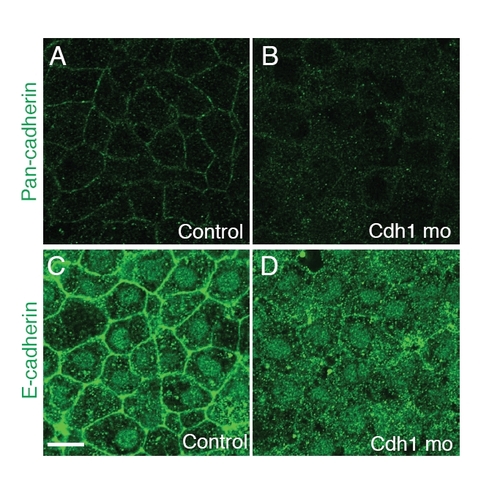
Cdh1 morphants have decreased staining with E-cadherin specific or pan-cadherin antibodies. Confocal images of the most external layer of deep cells at the animal pole of fixed embryos at 50% epiboly stained with a pan-cadherin antibody (A,B) and a zebrafish E-cadherin specific antibody (C,D). Control, uninjected WT; cdh1 mo, embryos injected with cdh1 morpholino. Scale bar: <10 μm. |
Expression Data
Expression Detail
Antibody Labeling
Phenotype Data
Phenotype Detail
Acknowledgments
This image is the copyrighted work of the attributed author or publisher, and
ZFIN has permission only to display this image to its users.
Additional permissions should be obtained from the applicable author or publisher of the image.
Full text @ Development