
FIGURE
Fig. 7
Fig. 7
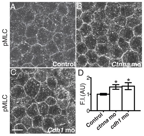
cdh1 and ctnna morphant DCs displayed increased phospho-MLC at cell contacts. (A-C) Confocal images of the epiblast most external layer at the animal pole of embryos at 50% epiboly stained for phospho-MLC. Scale bar: 10 μm. (D) Fluorescence intensity (FI) at cell contacts of embryos stained for pMLC. Three independent experiments, n=300 cell contacts (15 embryos) per experiment. *P<10–3 versus the control (normalized to 1). Error bars indicate s.d. |
Expression Data
Expression Detail
Antibody Labeling
Phenotype Data
Phenotype Detail
Acknowledgments
This image is the copyrighted work of the attributed author or publisher, and
ZFIN has permission only to display this image to its users.
Additional permissions should be obtained from the applicable author or publisher of the image.
Full text @ Development