
Fig. 3
- ID
- ZDB-FIG-111223-23
- Publication
- Herwig et al., 2011 - Distinct Cellular Mechanisms of Blood Vessel Fusion in the Zebrafish Embryo
- Other Figures
- All Figure Page
- Back to All Figure Page
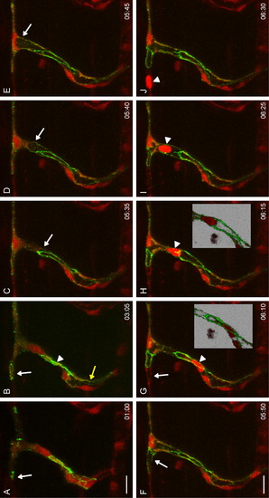
Anastomosis by Membrane Invagination Still pictures from Movie S3 showing a time-lapse of a Tg(fli:GAL4FFubs3; UAS:mRFPrk8; UAS:EGFP-ZO1ubs5) embryo (total length: 10:25). (A and B) A tip cell has formed initial contacts and subsequently forms loops of EGFP-ZO1 with its adjacent partner (white arrows). The stalk shows a multicellular organization (two lines of EGFP-ZO1, arrowhead in B) and an opening lumen (yellow arrow in B). (C) The lumen projects into the fusion cell (follow arrow from C to E) and finally reaches the left ring of EGFP-ZO1, which is subsequently inflated (arrow in F). The lumen then continues to extend into the unlabeled fusion cell on the left (arrow in G). A blood cell then passes through the newly formed lumen (follow arrowhead from G–J). Insets show the blood cell passing behind the junction (G) and in front of the junction (H). More examples of membrane invagination are shown in Movie S4, Movie S6, and Figures S3B and S3C. Scale bar in all pictures represents 20 μm. |