
Fig. 3
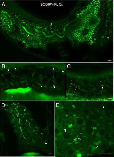
BODIPY-FL C5 reveals significant subcellular details within larval digestive organs (6dpf). A. In enterocytes of the anterior intestine BODIPY-FL C5 appears in structures reminiscent of lipid droplets. Lumenal accumulation of BODIPY-FL C5 in the intestine (arrow) is consistent with the expedited metabolism, transport and biliary concentration of medium chain fatty acids. The analog also appears in a dorsal intestinal blood vessel (arrowhead) and in the liver (asterisk). B. Subcellular enterocyte morphology is visualized following a BODIPY-FL C5 feed. Lipid droplets (filled arrowhead), a nucleus (empty arrowhead), apparent pericellular accumulation of the analog (arrows) and the intestinal lumen (asterisk) are indicated. C. BODIPY-FL C5 visualizes subcellular details of acinar cells and pancreatic ducts in the larval exocrine pancreas. A single acinar cell (dashed box) containing a basal nucleus and apical zymogen granules, as well as pancreatic ducts (arrowhead) is indicated. D. BODIPY-FL C5 feeding illuminates the intrahepatobiliary ductal network. In the right liver lobe, a single long duct (arrow), numerous interconnecting ducts and terminal ductules (arrowhead) are illuminated (see also Supplementary Movie-1). E. Subcellular details of larval hepatocytes are revealed by BODIPY-FL C5 feeding. Lipid droplets (filled arrowhead), hepatocyte nuclei (empty arrowhead), and intrahepatic ducts (arrow) are indicated. A. is a composite of 2 separate confocal images. Scale bars = 10μm. (n = 3; 6 larvae per feed.) BODIPY-FL C5 reveals significant subcellular details within larval digestive organs (6dpf). A. In enterocytes of the anterior intestine BODIPY-FL C5 appears in structures reminiscent of lipid droplets as well as in small endosomal-like compartments. Lumenal accumulation of BODIPY-FL C5 in the intestine (arrow) is consistent with the expedited metabolism, transport and biliary concentration of medium chain fatty acids. The analog also appears in a dorsal intestinal blood vessel (arrowhead) and in the liver (asterisk). B. Subcellular enterocyte morphology is visualized following a BODIPY-FL C5 feed. Lipid droplets (filled arrowhead), a nucleus (empty arrowhead), apparent pericellular accumulation of the analog (arrows) and the intestinal lumen (asterisk) are indicated. C. BODIPY-FL C5 visualizes subcellular details of acinar cells and pancreatic ducts in the larval exocrine pancreas. A single acinar cell (dashed box) containing a basal nucleus and apical zymogen granules, as well as pancreatic ducts (arrowhead) indicated. D. BODIPY-FL C5 feeding illuminates the intrahepatobiliary ductal network. In the right liver lobe, a single long duct (arrow), numerous interconnecting ducts and terminal ductules (arrowhead) are illuminated (see also Supplementary Movie-1). E. Subcellular details of larval hepatocytes are revealed by BODIPY-FL C5 feeding. Lipid droplets (filled arrowhead), hepatocyte nuclei (empty arrowhead), and intrahepatic ducts (arrow) are indicated. A. is a composite of 2 separate confocal images. Scale bars = 10μm. (n = 3; 6 larvae per feed.) |
Reprinted from Developmental Biology, 360(2), Carten, J.D., Bradford, M.K., and Farber, S., Visualizing digestive organ morphology and function using differential fatty acid metabolism in live zebrafish, 276-85, Copyright (2011) with permission from Elsevier. Full text @ Dev. Biol.