
Fig. S3
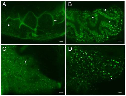
Egg yolk feeding enhances BODIPY-FL C16 uptake and lipid droplet formation in the larval intestine and liver. A. Zebrafish larvae (6 dpf) fed BODIPY-FL C16 in the absence of egg yolk exhibit fluorescence in the intestinal lumen (arrow) and faintly in enterocytes (arrowhead). B. Co-feeding BODIPY-FL C16 with egg yolk enhances analog uptake by intestinal enterocytes. Lipid droplets (arrowhead) appear in enterocytes surrounding the intestinal lumen (asterisk). Nuclei (arrow) appear as dark circles. C. Larvae fed BODIPY-FL C16 in the absence of egg yolk have diffuse accumulation of the analog in their livers. Few lipid droplets appear in hepatocytes, with fluorescence appearing mainly in the cytoplasm of hepatocytes and in hepatic ducts (arrow). D. The livers of larvae co-fed BODIPY-FL C16 with egg yolk have lipid droplets (arrow). Scale bars = 10μm (n = 3 feeds; 4 larvae imaged per feed.) |
Reprinted from Developmental Biology, 360(2), Carten, J.D., Bradford, M.K., and Farber, S., Visualizing digestive organ morphology and function using differential fatty acid metabolism in live zebrafish, 276-85, Copyright (2011) with permission from Elsevier. Full text @ Dev. Biol.