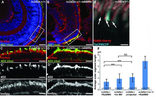
rab8 morphant phenotype and Rab8 localization at the base of the outer segment. (A-B) 4 dpf retinal cryosections of wild-type (A) and rab8 morphant (rab8MO)(B) embryos showing shortened outer segments stained with bodipy (red) and rhodopsin (4D2 antibody, green). (C-D) Higher power images of boxed areas in A and B with separate channel images for 4D2 (C′-D′) and bodipy (C′′-D′′) showing shortened and dysmorphic outer segments as well as rhodopsin mislocalization. (E) Rab8-cherry expression (red) in cryosections from wild-type TaCP:MCFP-expressing photoreceptors (outer segments are light blue) showing punctate localization at the base of the outer segment. Scale bars are 40 µm in A-B, 10 µm in C-D′′ and 4 µm in E. (F) Average outer segment length in cc2d2a -/- embryos injected with Rab8MO, injected with a control MO, uninjected and cc2d2a +/+ or +/- embryos injected with Rab8Mo. Scale bars are standard deviations. *** p<0.0001, NS non significant (p=0.7, Chi-square test). PR photoreceptors.
|
