
Fig. S15
- ID
- ZDB-FIG-111201-16
- Publication
- Mich et al., 2011 - Hedgehog and retinoic acid signaling cooperate to promote motoneurogenesis in zebrafish
- Other Figures
- All Figure Page
- Back to All Figure Page
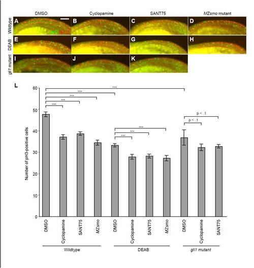
Hh signaling, basal Gli activity and RA signaling cooperate to promote cell proliferation in the anterior spinal cord. (A-K) Embryos cultured with (A,E,I) DMSO, (B,F,J) 100 μM cyclopamine, or (C,G,K) 100 μM SANT75, as well as (D,H) MZsmo mutants, were co-treated with (E-H) 10 µM DEAB. gli1 mutants were similarly treated with DMSO (I) or the Smoothened antagonists (J,K). The embryos were then fixed at the 18-somite stage and stained for phosphohistone H3 (pH3; green) to mark mitotic cells and Isl1/2 (red) to label the posterior neural tube. The Isl1/2-expressing cells were used to determine the dorsoventral and mediolateral positions of the neural tube, which can also be morphologically distinguished from overlying skin cells. Lateral views of the neural tube between approximately the first and eighth somites are shown, anterior to the left. The bright yellow fluorescence at the bottom of each micrograph is yolk autofluorescence. (L) Quantification of pH3-positive cells in the anterior neural tube between the first and eighth somites. Data are the average number of pH3-positive cells per embryo ± s.e.m. ***, P<0.001. Five embryos were analyzed for the gli1 mutant phenotypes, and at least 10 embryos were evaluated for all other conditions. Scale bar: 50 μm. |