
Fig. 4
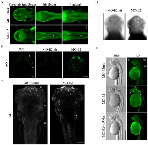
Confocal imaging analysis of hydrocephalus and apoptosis in lgi1b morphants. Representative single z-plane images (A) from (left) forebrain and midbrain, (center) hindbrain and (right) Z-stack images show MO-E2 morphants have enlarged ventricles (indicated by *) in both forebrain/midbrain and midbrain/hindbrain compared with mismatch controls (MO-E2mis). Scale bar = 50 μm. (B) Uninjected (WT), MO-E2mis and MO-E2 embryos stained with AO after 48 hpf showed increased cell death (green cells). (C) AO staining analysis showed increased apoptotic cells (White dots) could be detected in the forebrain of the lgi1b morphants after 72 hpf. (D) Whole mount TUNEL analysis further supports increased apoptosis in the forebrain at 72 hpf in lgi1b morphants. Black dots (white arrows) indicate TUNEL-positive cells. (E) AO staining shows apoptosis (arrows) was significantly reduced in lgi1b morphants co-injected with lgi1b mRNA at 36 hpf. (B–D) dorsal view, Z-stack confocal images. (E) a single lateral view image. Scale bars: B, C and D, 100 μm; E, 500 μm. |
| Fish: | |
|---|---|
| Knockdown Reagent: | |
| Observed In: | |
| Stage Range: | Prim-25 to Protruding-mouth |