
FIGURE
Fig. S4
- ID
- ZDB-FIG-110928-32
- Publication
- Culbertson et al., 2011 - Chondrogenic and Gliogenic Subpopulations of Neural Crest Play Distinct Roles during the Assembly of Epibranchial Ganglia
- Other Figures
- All Figure Page
- Back to All Figure Page
Fig. S4
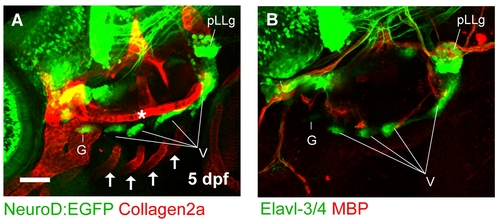
Spatial relationship between neurons, glia, and cartilage in the zebrafish head. (A) Lateral view of cranial ganglia in a 5 dpf wildtype larva expressing Tg(neurod:EGFP) and stained for Collagen2a (red). The rostral basicranial commisure (asterisk) can be seen positioned dorsally to the cranial ganglia. The Collagen2a-positive branchial arches are located ventrally (arrows). (B) Lateral view of cranial ganglia in a 5 dpf wildtype larva as visualized with immunofluoresence against Elavl-3/4 (green). Glial cells are stained with an antibody recognizing MBP (red). Scale bar in (A) = 50 μm. |
Expression Data
| Genes: | |
|---|---|
| Antibodies: | |
| Fish: | |
| Anatomical Terms: | |
| Stage: | Day 5 |
Expression Detail
Antibody Labeling
Phenotype Data
Phenotype Detail
Acknowledgments
This image is the copyrighted work of the attributed author or publisher, and
ZFIN has permission only to display this image to its users.
Additional permissions should be obtained from the applicable author or publisher of the image.
Full text @ PLoS One