
Fig. 6
- ID
- ZDB-FIG-110915-14
- Publication
- Zygmunt et al., 2011 - Semaphorin-PlexinD1 Signaling Limits Angiogenic Potential via the VEGF Decoy Receptor sFlt1
- Other Figures
- All Figure Page
- Back to All Figure Page
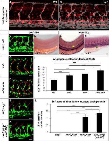
Notch Signaling Loss Does Not Phenocopy obd (A and B) Expression of Notch′s activity nuclear reporter Tg(Tp1bglob:hmgb1-mCherry)jh11 (red) in the endothelium (gray) of WT (A) and obd (B). (C–F) obd; mib. (C) Endothelium, green. SBs, red. (D–F) WISH with sflt1, flt4 and mflt1 riboprobes, as indicated. Double mutant phenotypes classed as obd-like (C and D) or mib-like (E and F) based on the mutant they resemble most. Note lack of sflt (as in Figure 3G) and ectopic aortic flt4 (yellow arrowhead; as in Figure S6A) and venous mflt1 stainings (green arrowhead, as in Figure S6B). (G–I) Angiogenic cell abundance within the trunk′s arterial tree of WT, obd, mib (G) and obd; mib (H) in Tg(fli1:nEGFP)y7 embryos. (G–H) EC nuclei, green. SBs, red. (I) Quantification; n = 10 per genotype. (J–L) SeA sprout abundance in plcg, mib; plcg, obd; plcg (J) and obd; plcg embryos injected with 10 ng of mib MO (mib MO) (K). (J and K) Endothelium, green. SBs, red. (L) n = 8, 7, 11 and 9 for plcg, mib; plcg, obd; plcg and obd; plcg (mib MO), respectively. Scale bars represent 50 μm (A, B, and D–F), 30 μm (C, G, H, J, and K). (I and L) *p < 0.05, ***p < 0.001. Error bars represent SEM. (A–F, G, H, J, and K) Anterior, left; dorsal, up. Trunk images and quantifications: 32 hpf (A–C, G–L), 28 hpf (D–F). |
| Gene: | |
|---|---|
| Fish: | |
| Anatomical Term: | |
| Stage: | Prim-15 |
| Fish: | |
|---|---|
| Knockdown Reagent: | |
| Observed In: | |
| Stage: | Prim-15 |
Reprinted from Developmental Cell, 21(2), Zygmunt, T., Gay, C.M., Blondelle, J., Singh, M.K., Flaherty, K.M., Means, P.C., Herwig, L., Krudewig, A., Belting, H.G., Affolter, M., Epstein, J.A., and Torres-Vazquez, J., Semaphorin-PlexinD1 Signaling Limits Angiogenic Potential via the VEGF Decoy Receptor sFlt1, 301-314, Copyright (2011) with permission from Elsevier. Full text @ Dev. Cell