
Fig. 5
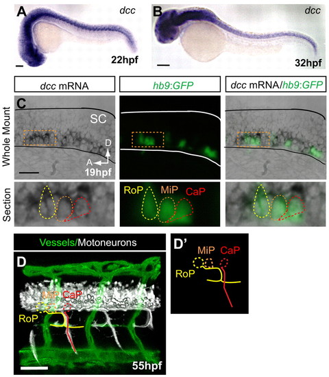
dcc is not detected in endothelial secondary sprouts but is expressed in motoneurons with axons present at the HMS prior to PAC formation. (A,B) dcc mRNA is expressed in the ventral spinal cord at 22 and 32 hpf and is not detectable in the secondary sprouts or other trunk vasculature. (C) Whole-mount and longitudinal section of hb9:GFP transgenic zebrafish embryo showing dcc mRNA ISH (purple), where hb9:GFP labels only motoneurons at early stages (green). (D) Lateral volume rendering of fli1a:dsRedEx; hb9:GFP embryo at 55 hpf. Double transgenic labels endothelial cells (green) and motoneuron axons (white) in the trunk. Rostral primary motoneuron (RoP, yellow) axons are present at the HMS adjacent to the PAC. (D2) Outline of motoneurons alone. CaP, caudal primary motoneuron (red); MiP, middle primary motoneuron (orange; axon omitted for clarity). SC, spinal cord; A, anterior; D, dorsal. Scale bars: 100 μm. |
| Genes: | |
|---|---|
| Fish: | |
| Anatomical Terms: | |
| Stage Range: | 20-25 somites to Long-pec |