
Fig. 1
- ID
- ZDB-FIG-110622-24
- Publication
- Waxman et al., 2011 - Zebrafish retinoic acid receptors function as context-dependent transcriptional activators
- Other Figures
- All Figure Page
- Back to All Figure Page
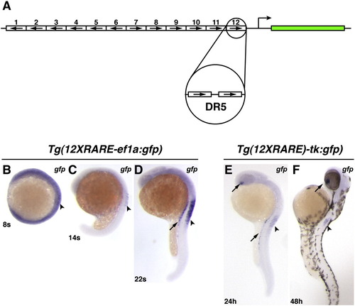
Zebrafish RARE transgenic reporter lines. (A) Schematic representation of the 12XRARE reporters. Arrows indicate direction of each DR5 RARE site. (B) At 8 somites, the expression of gfp from Tg(12XRARE-ef1a:gfp) can be seen at low levels in the anterior spinal cord (arrowhead). At this stage, embryos require a much longer exposure to visualize gfp expression than at subsequent stages. (C) At 14 somites, gfp expression is more easily detected in the spinal cord (arrowhead). (D) By 22 somites, the reporter is expressed at higher levels in the pronephros (arrow) and spinal cord (arrowhead). Shortly afterward, the reporter is also expressed in the ventral eye (not shown), similar to Tg(12XRARE-tk:gfp). (E) Expression of Tg(12XRARE-tk:gfp) does not initiate until closer to 24 hpf, when it is seen in the ventral eye (upper arrow), pronephros (lower arrow), and the anterior spinal cord (arrowhead). (F) Expression of Tg(12XRARE-tk:gfp) in the eye (arrow) and spinal cord (arrowhead) is maintained past 48 hpf. All images are lateral views, with anterior up and dorsal to the right. |
Reprinted from Developmental Biology, 352(1), Waxman, J.S., and Yelon, D., Zebrafish retinoic acid receptors function as context-dependent transcriptional activators, 128-140, Copyright (2011) with permission from Elsevier. Full text @ Dev. Biol.