|
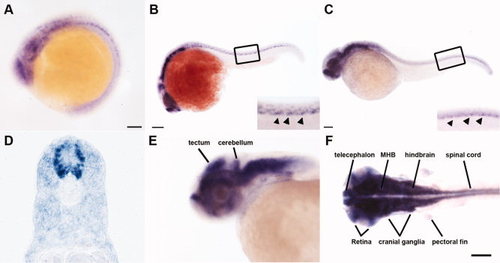
thsd7a transcripts were detected in the zebrafish developing nervous system. Representative embryos show the spatiotemporal expression pattern of thsd7a transcripts by in situ hybridization. Anterior is to the left. A: thsd7a expression was first detected at 15 hours postfertilization (hpf). B: At 22 hours postfertilization (hpf). The inset image is an enlarged view of the boxed region. The unique thsd7a expression pattern along the ventral edge of neural tube is indicated by arrowheads. C: At 32 hpf. Expression of thsd7a along the ventral edge of neural tube is indicated by arrowheads in the inset image enlarged from boxed region. D: Cross-section through the trunk region at 32 hpf. E,F: At 48 hpf, thsd7a is expressed in the midbrain, hindbrain, midbrain–hindbrain boundary (MHB), cerebellum, telencephalon, tectum, retina, cranial ganglia, spinal cord, and pectoral fin. F: Dorsal view. Scale bars = 20 μm in A; 50 μm in B; 100 μm in C,F.
|