
Fig. 4
- ID
- ZDB-FIG-110527-31
- Publication
- Hong et al., 2011 - The transcriptional mediator component med12 is required for hindbrain boundary formation
- Other Figures
- All Figure Page
- Back to All Figure Page
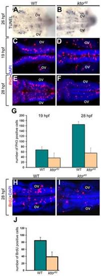
Cell proliferation defects in the hindbrain of ktoy82 mutants. All images are dorsal views at 19 hpf (C,D), 26 hpf (A,B), and 28 hpf (E,F,H,I). (A,B) Analysis of cell death using TUNEL assay. (C–G) PH3 staining (C–F), and quantification (G) of PH3 positive cells in wt and kto embryos. (H–J) BrdU staining of wt and mutant embryos (H,I), and quantification of BrdU positive cells (J). Five embryos were counted for each condition; the error bars indicate 1 standard deviation based on 5 samples. PH3 staining at 19 hpf, wt vs. kto: p = 0.003456; at 28 hpf, wt vs. kto: p = 2.97e-05. BrdU staining, wt vs. kto: p = 0.000706. ov, otic vesicle. |
| Fish: | |
|---|---|
| Observed In: | |
| Stage Range: | 20-25 somites to Prim-5 |