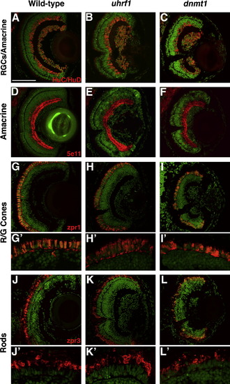
Fig. S4
- ID
- ZDB-FIG-110214-25
- Publication
- Tittle et al., 2011 - Uhrf1 and Dnmt1 are required for development and maintenance of the zebrafish lens
- Other Figures
- All Figure Page
- Back to All Figure Page
|
Differentiated retinal cell types are present in uhrf1 and dnmt1 mutants. Cryosections of eyes from wild-type (A,D,G,J), uhrf1 (B,E,H,K) and dnmt1 (C,F,I,L) eyes at 5 dpf. For all panels, antibody staining is in red and sections are counterstained with Sytox-Green (green) to stain nuclei. (A–C) HuC/HuD staining of retinal ganglion cells and amacrine cells. (D–F) 5e11 staining of amacrine cells. (G–I) zpr-1 staining of red/green cones. (J–L) zpr-3 staining of rods. Each of these differentiated cell types is present and in appropriate laminar positions in uhrf1 and dnmt1 mutant retinas. Despite correct localization, both red/green cones and rods have a distorted morphology in uhrf1 and dnmt1 mutant retinas (H′,I′,K′,L′), the severity of which correlated with the severity of lens phenotype. Scale bars are 80 μm. |
| Fish: | |
|---|---|
| Observed In: | |
| Stage: | Day 5 |
Reprinted from Developmental Biology, 350(1), Tittle, R.K., Sze, R., Ng, A., Nuckels, R.J., Swartz, M.E., Anderson, R.M., Bosch, J., Stainier, D.Y., Eberhart, J.K., and Gross, J.M., Uhrf1 and Dnmt1 are required for development and maintenance of the zebrafish lens, 50-63, Copyright (2011) with permission from Elsevier. Full text @ Dev. Biol.

