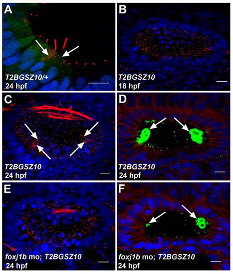
Foxj1b is required for the differentiation of motile and kinocilia. (A) Expression of GFP in the hair cells of a heterozygous T2BGSZ10 transgenic zebrafish embryo. Arrows indicate hair cells. (B) Defective motile cilia differentiation in the ear of a homozygous T2BGSZ10 transgenic embryo (compare with Fig. 1E). (C) Defective kinocilia differentiation in the ear of a homozygous T2BGSZ10 transgenic embryo. Note the variable lengths of the kinocilia (arrows) (compare with Fig. 1F). (D) Malformed otoliths (green, arrows) in the ear of a homozygous T2BGSZ10 transgenic embryo. (E) A foxj1b morpholino-injected homozygous T2BGSZ10 transgenic embryo showing a more pronounced effect on ciliary differentiation with severe reduction in the motile and kinocilia. (F) A more prominent effect on otolith (green, arrows) formation in the ear of a foxj1b morpholino-injected homozygous T2BGSZ10 transgenic embryo. Embryos in A-C,E were stained with anti-acetylated tubulin antibodies (red), and those in D and F with anti-Stm antibodies (green). The embryo in A was co-stained with anti-GFP antibodies (green), whereas those in D and F were co-stained with anti-β-catenin antibodies (red). Nuclei were visualized with DAPI (blue). All panels show lateral views of otic vesicles with anterior to the left. Scale bars: 10 μm.
|
